Graphene nanoribbons
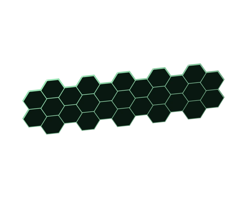
Cutting a graphene sheet leaves an atomic edge whose properties can be investigated through spectroscopy and calculations. The two views make armchair and zigzag terminations recognisable.
Formula and scientific context ↗Research background in physics
Cutting a graphene sheet leaves an atomic edge whose properties can be investigated through spectroscopy and calculations. The two views make armchair and zigzag terminations recognisable.
Formula and scientific context ↗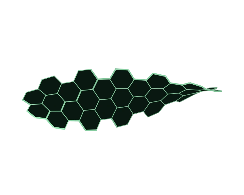
Twisting moves the hexagons out of plane and changes the orientation of ribbon regions, providing a visual introduction to the relationship between structural distortion and electronic properties.
Formula and scientific context ↗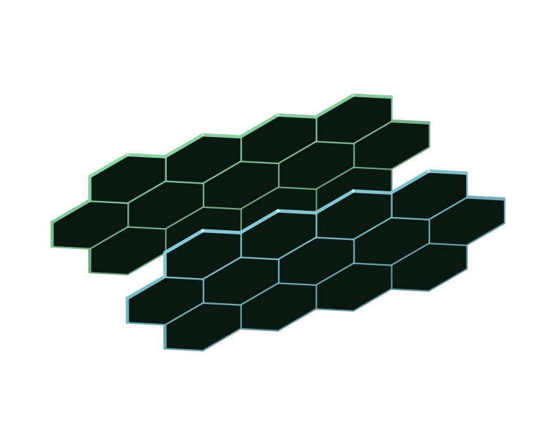
Two overlapping graphene fragments reveal how a relative shift changes ring alignment, introducing the role of interlayer interactions in the study of optical properties.
Formula and scientific context ↗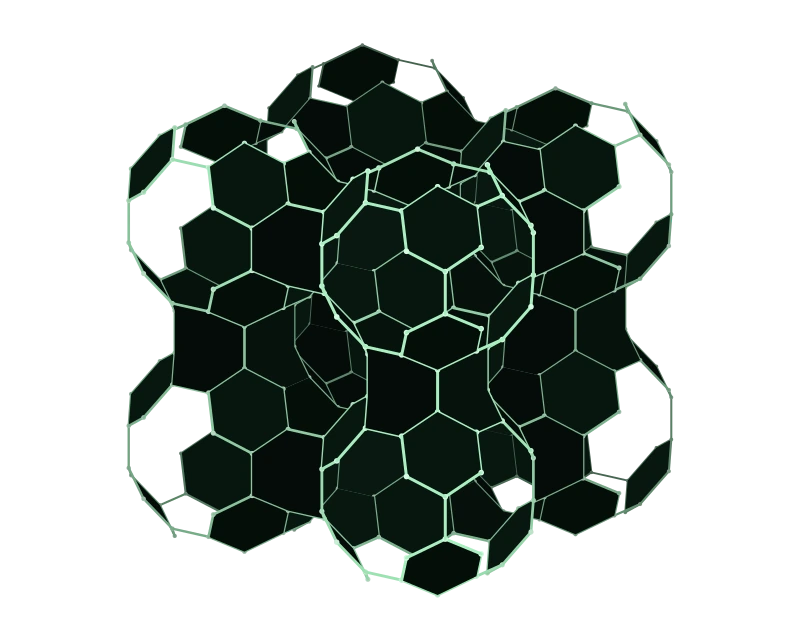
The carbon lattice's hexagons remain visible as the framework curves around its openings. Octagons connect the saddle regions, where the bonds reveal the narrowing and subsequent widening of the structure.
Formula and scientific context ↗Which properties of a nanostructure depend on its geometry and bond arrangement?
Graphene nanoribbons and nanoflakes, schwarzites and other carbon structures allow the study of how bond configurations affect a system’s electronic, optical and vibrational behaviour, while topological descriptions of negatively curved networks provide tools for relating structural organisation to the physical properties under consideration.
Ab initio calculations have been compared with spectroscopic results to identify Raman and vibrational signatures associated with nanoribbon edges. Other work examines how structural distortion changes the bandgap, the optical properties of coupled nanoflakes and the mathematical indices used to compare carbon networks.
PhD in Physics and Nanosciences: “Vibrational and optical fingerprints in graphene-based nanostructures” (2013–2016).
Research experience at CNR–NANO, Modena, on the optical properties of carbon nanostructures (2012–2013).
Atomic edges and vibrational signatures
Cutting a graphene sheet leaves an atomic edge whose properties can be investigated through spectroscopy and calculations. The two views make armchair and zigzag terminations recognisable.
Observe the atom pairs along the armchair edge, then select the zigzag view to compare its different boundary pattern. Follow the hexagons inward and examine the ends of the finite fragment.
Honeycomb lattice: two atoms per primitive cell.
Nearest neighbours: dᵢⱼ = 1 in model units.
Armchair and zigzag: two orientations of the same lattice.
The planar lattice has two atoms per primitive cell and unit bond lengths in this model. The cut direction determines an armchair or zigzag termination.
The finite fragments contain 80 and 84 carbon atoms respectively. Coordinates describe an ideal planar lattice; hydrogen and other terminating groups are omitted. The fragments have different finite dimensions and illustrate edge patterns. The linked Raman and HREELS papers examine specific structures through measurements and calculations. These figures introduce their geometry; peak assignments and frequencies belong to the papers’ experimental and theoretical results.
Lattice shape and electronic structure
Twisting moves the hexagons out of plane and changes the orientation of ribbon regions, providing a visual introduction to the relationship between structural distortion and electronic properties.
Follow an edge from one end to the other as it turns around the long axis. Compare central rings with those at the ends and rotate the model to distinguish the imposed twist from perspective.
R(x,y) = (x, y cos θ(x), y sin θ(x))
θ(x) = 0.16 x
The planar ribbon’s connectivity is preserved.
Each transverse coordinate rotates through an angle that increases linearly along the ribbon axis. The bond list remains identical to the planar ribbon.
This illustrative geometric deformation acts on 80 atoms. It preserves connectivity while changing some distances and bond angles. Its twist parameter is chosen for visual clarity. The 2018 paper examines specific ribbons and control of structural distortion; this figure introduces the question without claiming an optimised structure, a calculated bandgap or a normal vibrational mode.
Stacking and optical properties
Two overlapping graphene fragments reveal how a relative shift changes ring alignment, introducing the role of interlayer interactions in the study of optical properties.
Rotate the model to separate the two planes visually, then switch between shifted and aligned layers. Rings remain identical within each fragment while the relative positions of facing atoms change.
R₂ = R₁ + (Δx, 0, h)
h = 2.36 dCC; Δx = 1 dCC or 0
Two separate graphs, with no interlayer covalent bonds.
The second fragment is translated horizontally and separated vertically from the first. All drawn bonds belong to individual layers.
Two ideal 40-carbon fragments have a fixed separation and an illustrative relative shift. Hydrogen, functional groups and orbitals are omitted. Colours distinguish the layers. Aligned and shifted views preserve each fragment’s geometry. The linked paper derives optical results from calculations on specific systems; this figure depicts arrangement and overlap without simulating absorption or charge transfer.
Carbon and negative curvature
The carbon lattice's hexagons remain visible as the framework curves around its openings. Octagons connect the saddle regions, where the bonds reveal the narrowing and subsequent widening of the structure.
Follow the six-atom rings along the curved walls and identify the octagons connecting them through the neck. Rotate the lattice to follow repeating cavities in all three spatial directions, with hexagons outlining their curved walls and necks connecting adjacent cells. “Two curvatures” shows a blue axial section and an amber transverse section; at the pale point, they follow principal directions and bend in opposite senses. “P688 structure” broadens the view to the network of connected cavities.
cos(2πx) + cos(2πy) + cos(2πz) = 0
K = k₁ · k₂ < 0 in the displayed neck.
P688: cubic Im-3m cell, a = 7.828 Å, 48 atoms per cell.
The sum of the cosines of two pi times each coordinate is zero. Gaussian curvature K is the product of the principal curvatures k one and k two. Their opposite signs make the product negative in the neck.
The carbon positions follow the P688 crystallographic site (0.31952, 0.31952, 0.09373) and Im-3m symmetries; bonds join nearest neighbours at 1.452–1.467 Å. The initial view contains eight cells arranged 2×2×2, with 384 atoms, 64 complete hexagons and 48 complete octagons. The overview extends the periodic structure to 27 cells arranged 3×3×3, with 1,296 atoms. The finite boundaries cut rings and bonds that would continue through the periodic lattice. Dark ring faces distinguish front and rear bonds; they do not represent physical walls. The continuous surface appears in “Two curvatures” as a nodal guide for the Schwarz P family; its mean curvature can differ from zero, unlike the exact minimal surface. The carbon lattice and the reference surface are distinct descriptions. Colours, lighting and small offsets of the guide curves support legibility; carbon coordinates preserve the published model's distances, apart from uniform display scaling. This is a theoretical structure, with no new energy minimisation.